

In my recovery week 9 post, I had spoken to the surgeon about my progress and the limitation at the front of my ankle that I had been enduring for the better part of a year. This is the follow-up story to that problem.
April 8, 2025
Long before my Achilles ruptured, I had been trying to address an aggravating front-of-ankle limitation. When my Achilles blew out, suddenly, the ankle was no longer the focus of my rehabilitation.
As I started more weight-bearing and strength activity, I noticed the front of my ankle flaring up again. I told my PT about it, and at one point, the pain got really bad. He started including the ankle as part of our treatment sessions.
When I went to my last appointment with the surgeon, I mentioned that it seemed to be getting in the way of my Achilles progress. I kept hitting a wall with my exercises that had nothing to do with Achilles pain.
He decided it would be best to refer me to the foot and ankle specialist in the practice. He also suggested I wait a few more weeks into my recovery before I go. Whatever the result of that appointment might be, he wanted me to get further along into the Achilles healing process. If another surgery was indicated, that shouldn’t happen until I was more fully recovered.
The specialist visit
Two weeks later (recovery week 11), I returned to the office to see the new doctor. He was kind and professional. I gave him the full history of the front of ankle issue, how I ruptured the Achilles and how the ankle was impacting my Achilles progress. He did a manual exam, then suggested we start with an X-ray to rule out any mechanical issues with the bones. If there was nothing there, he’d prescribe an MRI to see what was happening in the soft tissue.
Looking at the possibilities, he said that a bone spur would likely warrant a surgery. Soft tissue, depending on what it was, could be addressed with cortisone first. If that didn’t work, then an arthroscopic “cleanout” could be an option a little further down the road. It all seemed reasonable to me. I just wanted answers.
The X-ray came back completely clean. No bone spurs, no arthritis (shockingly) and the joint spaces looked “perfect.” There were no anomalies in the bone structure that would be causing the pain.
An MRI was the next step. I scheduled it for about 3 weeks later.
The MRI
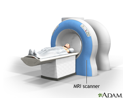
An MRI (magnetic resonance imaging) is a scan where the body part in question is rolled into a large donut-shaped apparatus.
You lay flat on a table, perfectly still, and the machine does its thing. Unfortunately, it takes about 20 minutes to complete the scan. During the process, it goes through phases of interminable banging, even through strong headphones to protect your ears. That’s the magnets whirring around the donut, which is what creates the images. It can be very unsettling and I had to breathe deep and focus on counting so I wouldn’t get too antsy on the table.
Before the scan, they ask you about 20 questions all relating to anything inside your body that might have metallic properties. If you do, they can’t do the scan. They would all be attracted to the magnetic forces and disaster could happen.
Fortunately for me, I have nothing implanted in my body. I was good to go. Also fortunate was the fact that I didn’t have to go in head first, since it was my ankle that needed scanning. I’ve had an MRI before for my shoulder and back where I went in head first for about 40 minutes. Not fun.
The results
The day after, I got the MRI report in MyChart. I’m always fascinated by medical imaging. I probably would have liked being in the medical field for that reason, but the sight of blood was an issue for me as a kid. But I digress…
The report was a bunch of medical jibber-jabber, and the impressions seemed a little daunting. This is what I saw:
- Edema-like marrow signal in the head and neck of the talus, with additional small subcortical changes of the anterior aspect of the distal tibia. Taken together, these findings may be seen in the setting of an anterior impingement syndrome.
- Areas of chondrosis at the talonavicular articulation and cartilage thinning at the talar dome. Additional small hypointense foci with peripheral rim of fluid talar dome and anterior aspect of the distal tibia, which may related to prior trauma.
- Tenosynovitis involving extensor digitorum longus and peroneal tendons, consistent with tenosynovitis. Additional tenosynovitis of flexor digitorum longus extending to the master knot of Henry, which may be seen in the setting of an intersection syndrome.
- Old injury of the anterior talofibular ligament, calcaneofibular ligament and deep fibers the deltoid ligament.
That’s a lot of medical jargon. As I read the details, I looked up every other word to create some context to understand what the heck it all meant. I shared the results with my PT and athletic trainer friend.
My PT told me to ask the doctor about a few things, including when he thought I should be able to do single heel raises. I had been struggling greatly with building the strength to push through them while standing.
My AT friend quick-texted me back: “On a quick reading, the results are that you are an athlete.” Ha ha. We spoke in greater detail before my doctor’s appointment and he helped me with things I needed to address with the doc.
An epiphany from the specialist
At the follow up appointment, I went to the visit armed with my list of questions. He asked how I was doing, and I replied, “well, based on the MRI results, it looks like my ankle is a giant mess.”
He smiled and said that it wasn’t as bad as it seemed. Yes, there was a little fluid buildup (edema), but just a little and he wasn’t too concerned about it. Yes, there were some old ligament tears, but since I wasn’t having any clinical issues of pair or loss of function, he wasn’t too concerned about that either. All of my tendons were intact with very limited fluid buildup. In general, things looked very good.
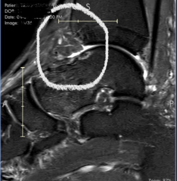
He opened up the images of the ankle and zoned in on the culprit.
At the front of the ankle joint, where the distal end of the tibia (the shin bone) meets the talus (the foot bone that connects to the tibia), there is some inflamed tissue that was invading the joint space. That is the reason why I felt pain and impingement whenever I flexed my foot and put weight into it. The bones kept hitting that soft tissue and couldn’t fully glide in it’s natural range of motion. The little white areas on the bone surface indicate some minor bone changes as a result.
Suddenly, everything made sense. Every time I took a long walking stride, stepped down from a higher level, or did a loaded calf stretch, it felt like something was getting in the way. That’s because there absolutely was something in the way.
He reiterated the treatment possibilities:
- Step one: cortisone shot to try calming down the inflammed tissue. I’d know in a few days if that was the answer.
- Step two: if the cortisone didn’t work, or didn’t last, he could go in arthroscopically, clean out the inflammed tissue, and hopefully regain my range of motion without the impingement.
To me, the answer was very clear. I asked him to do the cortisone shot, which he took care of right then and there, put a band aid on, and sent me on my way.
What about single calf raises?
Before I left, I asked him about the single heel raises. They were exceedingly difficult and I didn’t feel completely stable when doing them.
He suggested I lay off of the single heel raises for a while. Right now, I’m still in the early stages of healing the repair. Once I hit the six-month mark, I should be good to go. But right now, there’s still a significant danger of overstretching the repair. If it breaks, my problems increase exponentially. The double heel raises are fine to work now, since it does not require the repair to support my entire body weight. But for singles, it’s best to give it more time.
Message received, loud and clear.
Over the next few days, I paid close attention to how the ankle felt when I took long steps and descended the stairs. I didn’t want to make a premature assessment, but it seemed that the cortisone was doing something good. I went to the gym that Sunday and was mindful of how the ankle felt. For the most part, I did not feel any pinching or impingement like I had before. I’m sure my brain is expecting to feel it, but as I try to move more fluidly, I don’t have the physical barrier in the front anymore.
Time will tell if this fix is a permanent one. The cortisone could wear off and the tissue could get inflamed again, which would suck. But at least I know now what the problem is and the avenues to remedy it.

Very informative. I hope the cortisone shot helps. I’m dealing with two
similar problems in my right shoulder and right foot.Both are aggravated
doing my exercises for my leg. Just had the shots and am hoping they
will take care of the problems.
Iris
LikeLiked by 1 person